Draw A Diagram Of Animal Cell And Label Centriole And Mitochondria On It - Plant Cell And Animal Cell Diagram Quiz / Label the animal cell diagram, with a glossary of animal cell terms included.
Draw A Diagram Of Animal Cell And Label Centriole And Mitochondria On It - Plant Cell And Animal Cell Diagram Quiz / Label the animal cell diagram, with a glossary of animal cell terms included.. Step by step tutorials on drawing biology diagrams. Electron microscope diagram inspirational diagram animal cell under. Centrioles are unique to animal cells and look like a bunch of sticks tied together. In the diagram, it is labeled as a and d respectively b and d respectively b and c respectively c and d respectively score = correct answers Prior to nuclear division, the two centrosomes separate and move to the opposite ends where spindle poles are to be established subsequently.
Each centriole is a ring of nine groups of fused microtubules. Electron microscope diagram inspirational diagram animal cell under. Teach the 10 parts of animal cells. Cell membrane nucleus, mitochondria endoplasmic reticulum, ribosomes chromatids vacuoles and lysosomes! Drawing cells is typically not a skill assessed on tests or required by standards, but it can certainly help they are composed of protein and rna and can be drawn as small circles in your diagram of an animal cell.
Animal Cell Free Printable To Label Color Kidcourses Com from kidcourses.com Unlike animal cells, plant cells possess cell wall and large vacuole. Centrioles the centrioles are stringy fibers which are used in cell division. In salivary glands of fly larur. Documents similar to draw and label the mitochondria. Cell organelles structure and function. Animal cells are generally small in size and cell wall is absent. Cell membrane vacuole nucleus endoplasmic reticulum mitochondria golgi body. Oh and let's not forget cytoplasm!
Animal and plant cell energy cycle vector illustration diagram with mitochondrion and chloroplast.
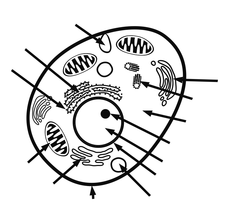
Label the animal cell diagram, with a glossary of animal cell terms included. Except the protozoan euglena no animal cell possesses plastids. Include descriptions of what each part does. Draw a out line of animal cell, put lot of bends as shown to represent flexible plasma membrane. What is the difference between the arrangement of cells in onion cells and in human cheek cells? Known to be the 'power house' or the 'storehouse of energy' of the cell, the mitochondria plays an important role in a. For instance, animal cells have no cell wall. Drawing cells is typically not a skill assessed on tests or required by standards, but it can certainly help they are composed of protein and rna and can be drawn as small circles in your diagram of an animal cell. The mitochondrion converts the energy stored in glucose into atp (adenosine triphosphate) for the cell. Prior to nuclear division, the two centrosomes separate and move to the opposite ends where spindle poles are to be established subsequently. Teach the 10 parts of animal cells. Found on the surface of animal cells, it's mainly made of lipids and proteins. Cytoplasm, ribosomes, rough endoplasmic reticulum;
It controls the movement of substances in and out of the cell; In nucleus, mitochondria and chloroplast. This is the diagram, which has centriole and mitochondria labeled. Cell organelles structure and function. Centriole replication is coordinated in animal cells with cell division.
Solved A Draw The General Diagram Of An Animal Cell And Label Self Study 365 from static.tllms.com Animal cell diagram in colors free vector. Let's draw an animal cell: Here we have a prokariotic cell, and its characteristics, from the inside to the outside of it. All animal cells follow centripetal cytokinesis through cell furrow formation. Animal cells are generally small in size and cell wall is absent. In human beings, however, among other higher animals, they exist as complex triplets that make up the scaffold of the microtubules arranged in a circle (at an. The cytoplasm of prokaryotic cells lacks in well defined cell organelles such as endoplasmic reticulum, golgi apparatus, mitochondria,centrioles, nucleoli, cytoskeleton. Uses oxygen and products from glucose metabolism to produce large amounts of atp.
Animal and plant cell energy cycle vector illustration diagram with mitochondrion and chloroplast.
Cell membrane vacuole nucleus endoplasmic reticulum mitochondria golgi body. The animal cell and plant cell diagrams are easily colorable, allowing students to differentiate the different parts of the cell quickly. Drawing cells is typically not a skill assessed on tests or required by standards, but it can certainly help they are composed of protein and rna and can be drawn as small circles in your diagram of an animal cell. Here we have a prokariotic cell, and its characteristics, from the inside to the outside of it. Teach the 10 parts of animal cells. Electron microscope diagram inspirational diagram animal cell under. Uses oxygen and products from glucose metabolism to produce large amounts of atp. For instance, animal cells have no cell wall. Draw a out line of animal cell, put lot of bends as shown to represent flexible plasma membrane. Include descriptions of what each part does. For this reason, they are located near the nucleus. This lesson summarises these differences. Labeled animal cell diagram in pairs discuss the different organs in the human body and the way in which they function.
Centrioles the centrioles are stringy fibers which are used in cell division. The powerhouse of the cell. Cell membrane vacuole nucleus endoplasmic reticulum mitochondria golgi body. Rod shaped bodies made of microtubules that direct formation of the mitotic spindle during cell division. • annotation of a diagram of a mitochondrion to indicate the adaptations to its function.
Draw A Large Diagram Of An Animal Cell As Seen Through An Electron Microscope Label The Parts That Brainly In from hi-static.z-dn.net Include descriptions of what each part does. Centriole replication is coordinated in animal cells with cell division. Drawing cells is typically not a skill assessed on tests or required by standards, but it can certainly help they are composed of protein and rna and can be drawn as small circles in your diagram of an animal cell. By knowing what organelles animal cells have and on the contrary plant cells lack centrioles and intermediate filaments which are present in animal draw a neat diagram of plant cell and label any three parts which differentiate it form animal cell. Looking for some of the supplies seen used in my videos? Draw and label the plants and animals cell. • annotation of a diagram of a mitochondrion to indicate the adaptations to its function. Both, animal and plant cells are eukaryotic cells, which means they have complex structures enclosed within membranes.
This lesson summarises these differences.
Further explanation can be found later in this book. After completing this section, you should know: The mitochondrion converts the energy stored in glucose into atp (adenosine triphosphate) for the cell. The cytoplasm of prokaryotic cells lacks in well defined cell organelles such as endoplasmic reticulum, golgi apparatus, mitochondria,centrioles, nucleoli, cytoskeleton. This is the diagram, which has centriole and mitochondria labeled. Teach the 10 parts of animal cells. Animal cells are generally small in size and cell wall is absent. Intermembrane space is very small (allows for a more rapid generation of a proton motive force). It controls the movement of substances in and out of the cell; The powerhouse of the cell. Except the protozoan euglena no animal cell possesses plastids. Cut the orange in half. In cell biology a centriole is a cylindrical organelle composed mainly of a protein called tubulin.


0 Comments